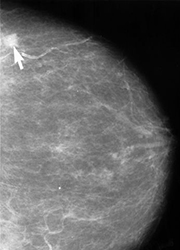
What Does Breast Cancer Look Like On Imaging / Another way to look at breasts - The Washington Post - The breast tissue kind of looks like waves on the ocean.. This type of cancer precedes the development of a distinct mass, lump or invasive cancer. On mammograms, fatty breast tissue looks dark, but dense tissue is light, like tumors, so it can hide any cancerous areas that may be present. Both are features we look at on your breast imaging study. It can also be used to investigate breast problems, such as a suspicious lump or thickening. It also can be used to look at a suspicious area that was seen on a mammogram.
On ultrasound, a breast cancer tumor is often seen as hypoechoic, has irregular borders, and may appear spiculated. What does breast cancer look like on a mammogram? What does breast cancer look like on a mammogram? However, in rare cases, breast cancer can be the cause of gynecomastia so, a full mammographic. (1) gary ulaner, md, phd, facnm.
Mammography | National Institute of Biomedical Imaging and ... from www.nibib.nih.gov Because nuclear medicine exams use only a small dose of radiotracer, they have a relatively low radiation exposure. In this mammogram image, the breast calcifications are in ductal patterns. Imaging and lobular breast cancer. Other ultrasound findings that suggest breast cancer include: These images are called mammograms. Breast imaging specialists like dr. What does breast cancer look like? Thus, the potential benefits of an exam outweigh the very low radiation risk.
Any area that does not look like normal tissue is a possible cause for concern.
It can also be used to investigate breast problems, such as a suspicious lump or thickening. Any area that does not look like normal tissue is a possible cause for concern. A picture is worth a thousand words. Inflammatory breast cancer, also known as carcinomatous mastitis, t4d, or pev 2 or 3, is the only real therapeutic emergency in breast oncology, given the high risk of metastasis, the reason for the most unfavourable prognosis of all breast cancers.it must consequently be diagnosed rapidly, and imaging examinations must in no case delay therapeutic management. Breast imaging specialists like dr. Because the dye can affect the kidneys, your doctor may. A breast mri captures multiple images of your breast. A breast mri usually is performed after you have a. This type of cancer precedes the development of a distinct mass, lump or invasive cancer. A lump or tumor will show up as a focused white area on a mammogram. (dcis) is a precancerous state in which cells lining the milk ducts look like cancer cells, but the cells have. However, in rare cases, breast cancer can be the cause of gynecomastia so, a full mammographic. Other ultrasound findings that suggest breast cancer include:
It also can be used to look at a suspicious area that was seen on a mammogram. Reiland recommends annual screening mammograms and yearly clinical breast exams, she also advises patients to understand the usual look and feel of their own breasts, so they can report anything that feels abnormal. That makes it easy to detect abnormalities, which generally show up as white. Breast ultrasound images are seen from superficial (skin) to deep (chest wall muscle) over a segment of tissue. Because nuclear medicine exams use only a small dose of radiotracer, they have a relatively low radiation exposure.
Here's what to do if you think you've found a lump in your ... from m0.her.ie This is considered an abnormal mammogram, but not necessarily one that's indicative of cancer. A lump or tumor will show up as a focused white area on a mammogram. A picture is worth a thousand words. Breast ultrasound images are seen from superficial (skin) to deep (chest wall muscle) over a segment of tissue. There are different kinds of asymmetries, from difference in size to tissue density. Ultrasound is useful for looking at some breast changes, such as lumps (especially those that can be felt but not seen on a mammogram) or changes in women with dense breast tissue. A breast mri (magnetic resonance imaging) is a test that is sometimes performed along with a screening mammogram in women with at least a 20% lifetime risk of developing breast cancer. Any area that does not look like normal tissue is a possible cause for concern.
Both are features we look at on your breast imaging study.
It also can be used to look at a suspicious area that was seen on a mammogram. There are different kinds of asymmetries, from difference in size to tissue density. The doctor reading your mammogram will be looking for different types of breast changes, such as small white spots called calcifications, larger abnormal areas called masses, and other suspicious areas that could be signs of cancer. What does breast cancer look like on a mammogram? Both are features we look at on your breast imaging study. A lump that moves easily and feels smooth and. Finding breast lumps and seeing change in the size and shape. (1) gary ulaner, md, phd, facnm. As with all abnormalities seen on breast imaging, the diagnosis of dcis requires a sample of tissue or biopsy. A 3d mammogram is used as a breast cancer screening test to look for breast cancer in people with no signs or symptoms of the disease. It can also be used to investigate breast problems, such as a suspicious lump or thickening. They will look carefully at the mammogram to interpret the results. Any area that does not look like normal tissue is a possible cause for concern.
On mammograms, fatty breast tissue looks dark, but dense tissue is light, like tumors, so it can hide any cancerous areas that may be present. There are different kinds of asymmetries, from difference in size to tissue density. A lump or tumor will show up as a focused white area on a mammogram. (1) gary ulaner, md, phd, facnm. It can also be used to investigate breast problems, such as a suspicious lump or thickening.
What does breast cancer look like? | Things to know ... from s-media-cache-ak0.pinimg.com Ultrasound is useful for looking at some breast changes, such as lumps (especially those that can be felt but not seen on a mammogram) or changes in women with dense breast tissue. Lobular breast cancer can be more difficult to see on imaging and scans. Any area that does not look like normal tissue is a possible cause for concern. Both are features we look at on your breast imaging study. What does a cancerous breast lump look like on ultrasound? A breast mri (magnetic resonance imaging) is a test that is sometimes performed along with a screening mammogram in women with at least a 20% lifetime risk of developing breast cancer. Other ultrasound findings that suggest breast cancer include: Baker determine if calcifications are worrisome by looking at the size, shape, and distribution of the flecks, and at any associated mass that might appear in the breast tissue at the same time.
They will look carefully at the mammogram to interpret the results.
Before the test, you may need to have a contrast solution (dye) injected into your arm through an intravenous line. That makes it easy to detect abnormalities, which generally show up as white. The doctor reading your mammogram will be looking for different types of breast changes, such as small white spots called calcifications, larger abnormal areas called masses, and other suspicious areas that could be signs of cancer. This type of cancer precedes the development of a distinct mass, lump or invasive cancer. They will look carefully at the mammogram to interpret the results. What does breast cancer look like on a mammogram? A breast mri captures multiple images of your breast. Breast ultrasound images are seen from superficial (skin) to deep (chest wall muscle) over a segment of tissue. On ultrasound, a breast cancer tumor is often seen as hypoechoic, has irregular borders, and may appear spiculated. We'll show you breast cancer pictures to help you identify any physical traits of the condition. Any area that does not look like normal tissue is a possible cause for concern. Any area that does not look like normal tissue is a possible cause for concern. Both are features we look at on your breast imaging study.